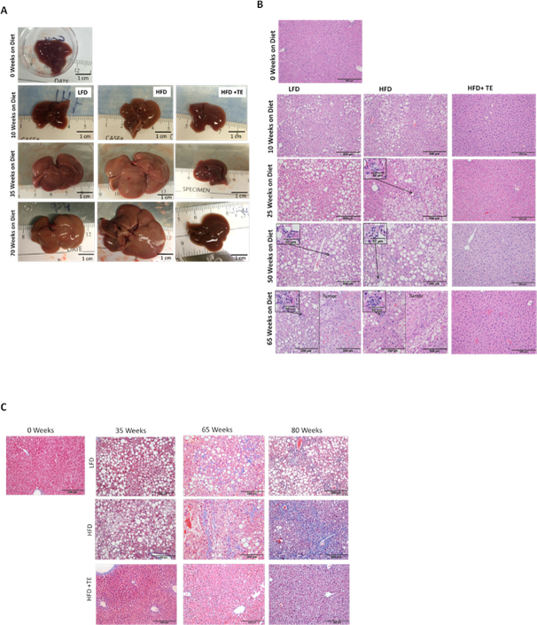
Figure 2. Comparison of liver morphology and histology among the groups.

(A) The gross morphology of representative non-tumor livers from 0, 10, 35 and 70 weeks on the diet. (B) H&E staining of livers at 0, 10, 35, 50 and 70 weeks on the diet (20X, scale bar indicates 200 μm). NASH found in livers of mice from 25 weeks and 50 weeks from HFD and LFD group, respectively, is shown in the Inlets. (C) Masson’s trichrome staining of representative sections at 0, 35, 65 and 80 weeks on diet. Blue staining indicates locations of expanded fibrotic tissue. Scale bar indicates 200 μm.
