Figure 5.

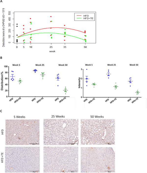
(A) γ-OHPdG levels in hepatic DNA measured by LC-MS/MS in mice fed HFD and HFD+TE for 0, 5, 10, 25, 35 and 50 weeks. The Wilcoxon rank sum test shows that the detectible levels of γ-OHPdG in HFD+TE group are significantly lower than that in HFD group (p=0.03) at week 50. (B) Quantification of γ-OHPdG by IHC staining in mouse livers from HFD vs. HFD+TE groups based on distribution (% of positively stained cells) and intensity (0 to 3) at week 5 (p=0.09 and p=0.12, respectively), week 25 (p=0.03 and p=0.0002, respectively) and week 50 (p=0.0016 and p=0.0006, respectively). In HFD mice, an increase of adduct levels from 5 to 25 weeks (p=0.0478 distribution and p=0.1699 intensity), followed by a decline at Week 50 (p=0.0190 and p= 0.0095, respectively). In HFD+TE mice, an increase from 5 to 25 weeks (p=0.0041 distribution), followed by decrease at Week 50 (p<0.0001 and p=0.0023, respectively) (C) Representative IHC staining of livers obtained from mice fed HFD vs. HFD+TE at weeks 5, 25 and 50.
