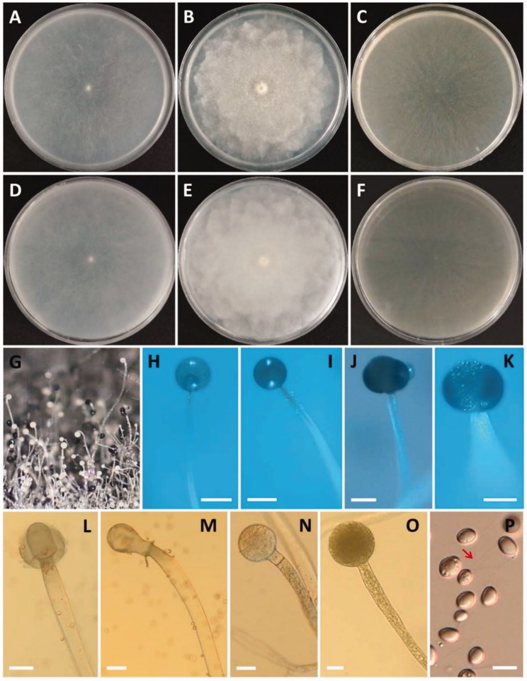
Figure 4.
Morphology of Gilbertella persicaria CNUFC-GWD3-9. (A, D) Colony on synthetic mucor agar; (B, E) Colony on potato dextrose agar; (C, F) Colony on malt extract agar (A–C, top view; D–F, reverse view); (G–I, N, O) Immature and mature sporangia and sporangiophores; (J, K) Wall suturing in two equal halves; (L, M) Columellae with collarette; (P) Sporangiospore with appendage (red arrow) (scale bars H, I = 200 μm; J–M = 50 μm; N, O = 20 μm; P = 10 μm).

