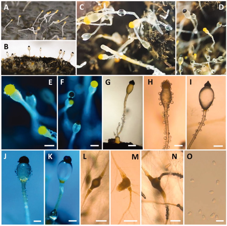
Figure 5.
Morphology of Pilobolus crystallinus CNUFC-EGF1-4. (A) Young sporangia and sporangiophores on dung agar medium; (B–K) Yellow and black sporangia, subsporangial vesicles, and sporangiophores (B–G, J, K, on water deer dung); (L–N) Substrate mycelia with trophocysts and rhizoidal extensions; (O) Sporangiospores (scale bars E–N = 200 μm; O = 10 μm).

