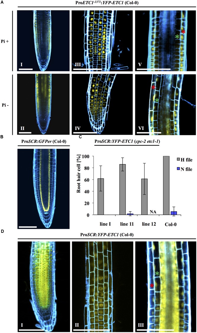
FIGURE 5.

Intercellular motility and localization of YFP-ETC1 under the ETC1 and SCR promoters. (A) Root of a cpc-2 etc1-1 double mutant plant carrying ProETC1:YFP-ETC1 under Pi+ and Pi– conditions. Under Pi+ conditions, YFP-ETC1 is seen in the root hair and non-root hair file epidermal cells (I, III) but not in the ground tissue (V). Under Pi– conditions, YFP-ETC1 was observed in the root hair and non-root hair file epidermal cells (II, IV) and in the ground tissue (VI). (I, II) Pictures show overview, (III, IV) pictures show focal planes of the epidermis and (V, VI) pictures show the focal planes in the cortical cells and the stele. Red stars mark epidermal cells, green stars mark cortical cells. Scale bar = 100 μm. (B) CLSM pictures of roots from transgenic ProSCR:GFPer (Col-0) plants grown under Pi+ conditions. Blue: propidium iodide, yellow: YFP. (C) A diagram showing the rescuing ability of ProSCR:YFP-ETC1 in the cpc-2 etc1-1 double mutant under Pi+ conditions. (D) ProSCR:YFP-ETC1 (Pi+ conditions). (I) Overview shows YFP-ETC1 in all tissues. (II) Focal plane showing YFP-ETC1 in epidermal cells. (III) Focal plane showing YFP-ETC1 in the epidermal cells and the ground tissue. Red star labels an epidermal cell, green star labels a cortical cell. Scale bar = 100 μm.
