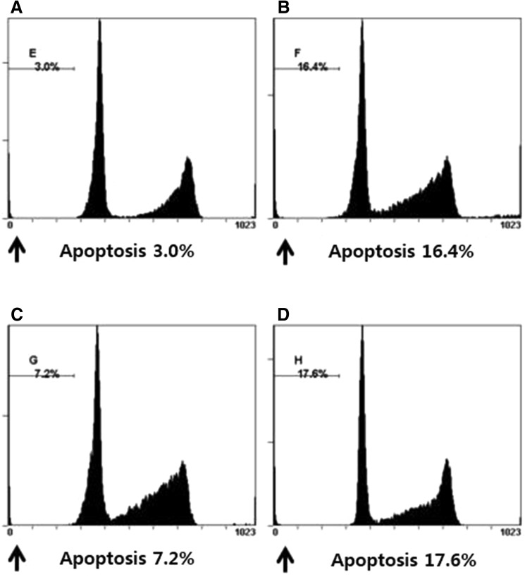
Fig. 5.
Flow cytometric assay was used to detect the percentage of apoptotic cells. Detection of apoptosis and propidium iodide staining. Every group of cells with propidium iodide staining was measured by flow cytometry. A The proportion of apoptosis cells in control group. B The proportion of apoptosis cells in H2O2 group. C The proportion of apoptosis cells in RPC/H2O2 group. D The proportion of apoptosis cells in 3-MA/RPC/H2O2 group

