
Fig. 1.
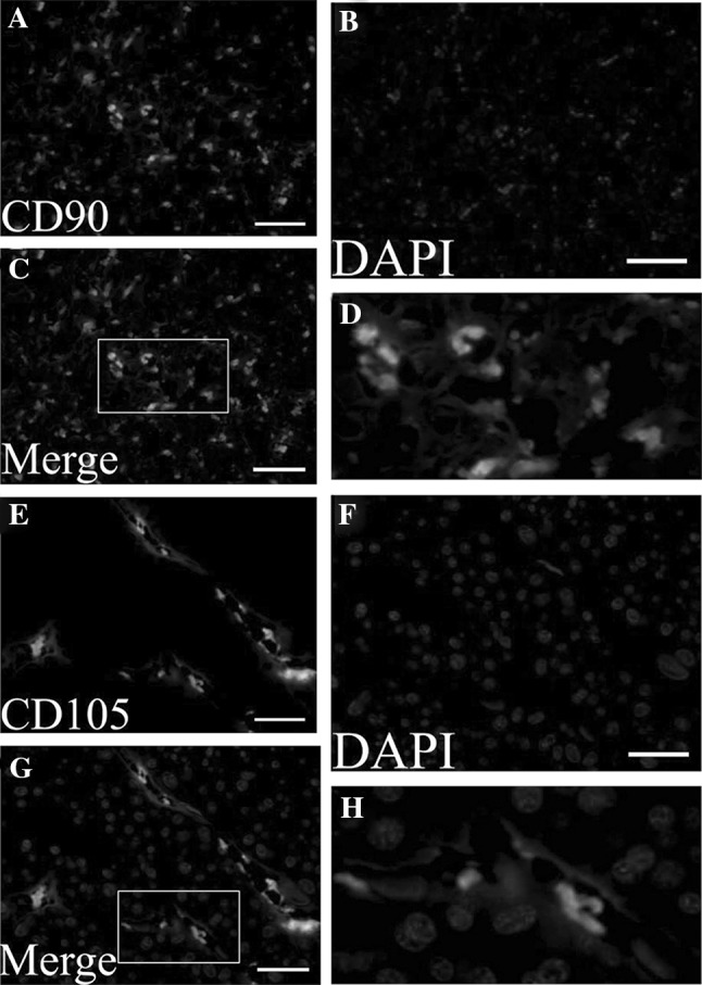
Result of mesenchymal stem cell surface markers positive in chorionic membranes with immunofluorescence staining. A and E show MSCs were stained with anti-CD90 (green) and anti-CD105 (green) antibodies. B and F show nuclei were stained with DAPI (blue). C and G show the result of merge with blue and green. D and H show the magnification display of selected rectangular region within C and G. Scale bar 100 μm. MSCs: mesenchymal stem cells. (Color figure online)