
Fig. 6.
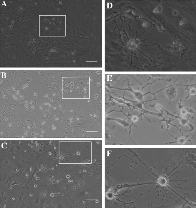
Morphological changes of the hCMCS were noticeable during induction of neuronal differentiation. Upon stimulation by neuronal differentiation medium 3 days (A), 7 days (B) and 14 days (C), spindle-shaped cells changed into neuron-like cells, as identified via microscopy. D, E and F show the magnification display of selected rectangular region within A, B and C. Scale bars 100 μm. hCMSCs: human chorionic membranes mesenchymal stem cells