
Fig. 7.
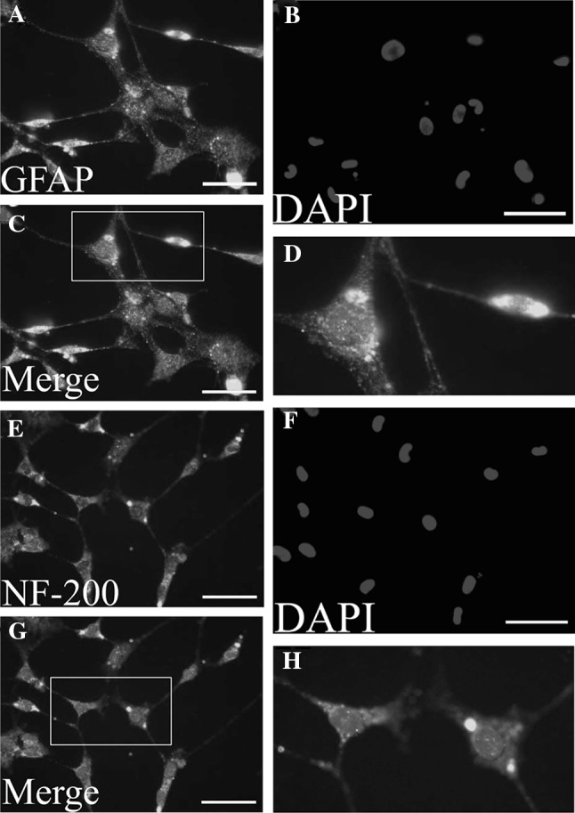
The hCMSCs were fixed and stained with antibodies against the GFAP (up panel) and NF-200 (down panel), and visualized under fluorescent microscopy. A and E show MSCs were stained with anti-GFAP (green) and anti-NF-200 (green) antibodies. B and F show nuclei were stained with DAPI (blue). C and G show the result of merge with blue and green. D and H show the magnification display of selected rectangular region within C and G. Scale bars 100 μm. hCMSCs: human chorionic membranes mesenchymal stem cells, GFAP: glial fibrillary acidic protein, NF-200: Neurofilament 200