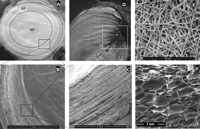
Fig. 1.
The AF and NP structure of an intervertebral disc in different magnifications (A, B, C), black squares show the magnified region. White square shows the NP extrusion through the AF (D), the place needs to be carefully focused for the AF treatment. Images “E” and “F” show two different scaffolds (E: nano-fibres and F: silk-base porous structure) that may be used for the torn AF re-construction

