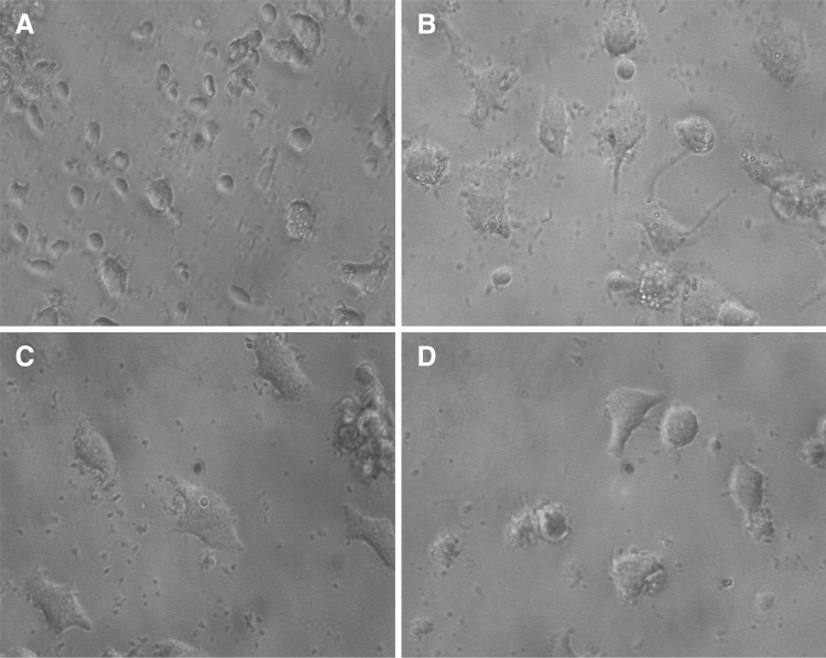
Fig. 7.
The effect of LPS, AMP and DEX on macrophage phenotype. Adherent peripheral blood-derived macrophages cultured for 3 days in the presence or absence of stimulators. Morphology of untreated A, LPS treated B, AMP treated C and DEX treated D macrophages was assessed by phase-contrast microscopy (×40 objective)

