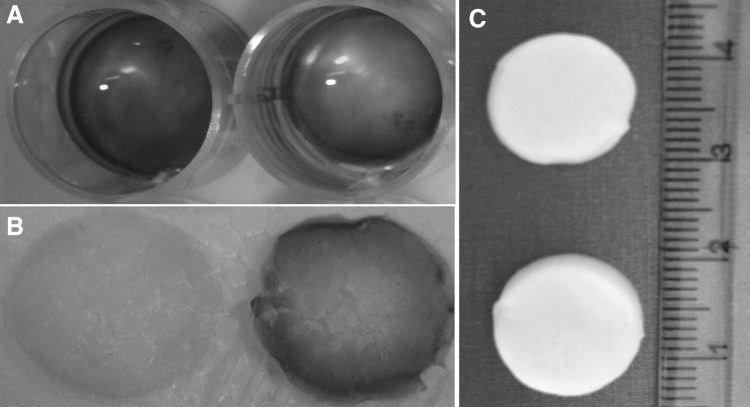
Fig. 1.
The gross structure of the gel and scaffold. The heparin immobilization was shown by toluidine blue staining. The heparinized gel A and scaffold B were stained purple (metachromatic) while the non-heparinized one stained blue (orthochromatic). The gross structure of the lyophilized scaffolds was also shown C

