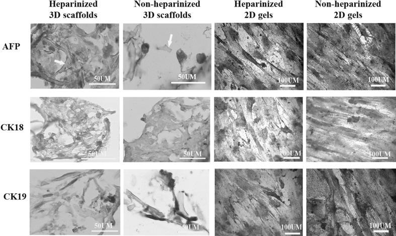
Fig. 5.
Immunohistochemistry and cytochemisrty of the cells cultured in 3D scaffolds and 2D gels in heparinized and non-heparinized conditions for alpha-fetoprotein (AFP, at the top), cytokeratin 18 (CL18, at the middle) and cytokeratin 19 (CK19, at the bottom). The cytoplasm of the differentiated cells was stained brown and the nuclei of undifferentiated cells stained with hematoxylin but the cytoplasm remained unstained. The arrows point collagen fibers in the scaffolds

