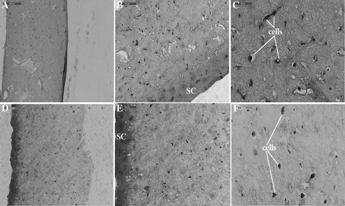
Fig. 6.
Microscopic images of stained histological sections of the hydrogels populated with primary fibroblasts (A–C gellan-crosslinked hydrogels at 10×, 20× and 40× objective magnifications; D–F pullulan-crosslinked hydrogels at the 10×, 20× and 40× objective magnifications). SC surface contraction of the hydrogel

