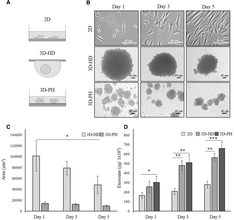
Fig. 1.
The effect of 3D culture on the exosome secretion from hBM-MSCs. A Schematic illustration of the experimental groups. hBM-MSCs were cultured in a monolayer at normal density (2D) or as 3D spheroids (3D-HD) at high cell density using the hanging-drop method or with a poly-HEMA coating (3D-PH), as described in Materials and Methods. B Microscopic morphology of MSCs in 2D, 3D-HD, and 3D-PH cultures after 1, 3, and 5 days. Scale bar: 60 μm. C Sizes of 3D MSCs spheroids cultured in 3D-HD and 3D-PH for 1, 3, and 5 days. The cross-sectional area of the spheroids was quantified from the bright-field images using Image J software. D The production efficiency of exosomes from MSCs cultured in 2D, 3D-HD, and 3D-PH. The amount of exosomes obtained from 1 × 10⁵ cells at 1, 3, and 5 days are shown. Data are presented as the mean and SD from three independent experiments in C and D. *p < 0.05, **p < 0.01 and ***p < 0.001 by one-way ANOVA. **** 3D: three-dimensional, hBM-MSC: human bone marrow-mesenchymal stem cells, 2D: two-dimensional, 3D-HD: 3D spheroids formed by the hanging drop method, 3D-PH: 3D spheroids formed by the poly-HEMA coating method, ANOVA: analysis of variance

