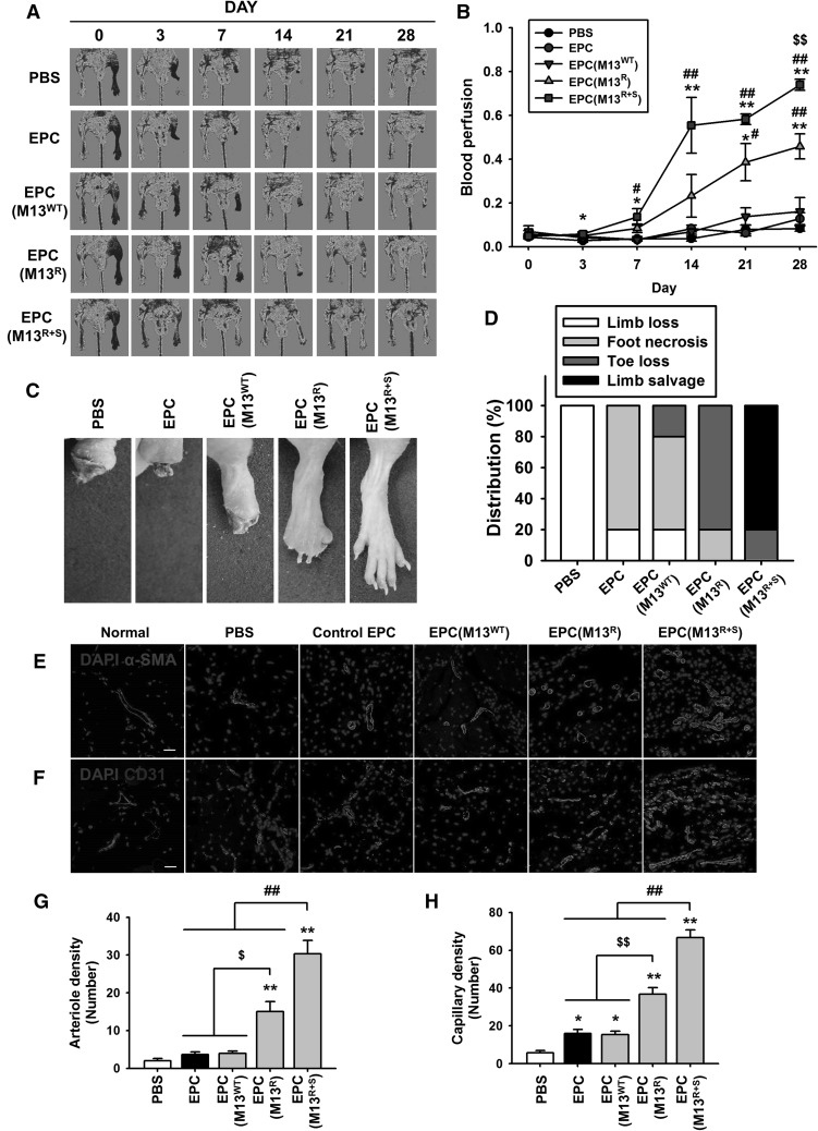
Fig. 5.
Assessment of the functional recovery in the murine hindlimb ischemia model. A The murine hindlimb ischemia model was established using Balb/C nude mice. The ratio of blood perfusion was assessed by laser Doppler perfusion imaging analysis in the ischemic limbs of the mice injected with PBS, untreated EPCs (EPC), EPCs treated with the wild type M13 phage (EPC (M13WT)), EPCs treated with M13 phage displaying RGD only (EPC (M13R)), and EPCs treated with M13 phage displaying both RGD and SDKP (EPC (M13R+S)) at 0, 3, 7, 14, 21, and 28 days post-surgery. B The ratio of blood perfusion was measured, and values are expressed as the mean ± SEM. *p < 0.05, **p < 0.01 versus PBS, #p < 0.05, ##p < 0.01 versus EPC (M13WT), and $$p < 0.01 versus EPC (M13R+S). C Representative images illustrating the various outcomes (limb loss, foot necrosis, toe lose, and limb salvage) of the ischemic limbs at post-operative day 28. D Distribution of the different outcomes at postoperative day 28. E, F At 28 days post-operation, the ischemic limb tissues were analyzed for vessel regeneration at the injured sites. Vessel formation was investigated by immunofluorescent staining for α-SMA(E) (arteriole density, green) and CD31(F) (capillary density, green). Scale bar = 20 μm. G, H Standard quantification of arteriole density (G) and capillary density (H) represented as the number of α-SMA- and CD31-positive cells per high-power field. Values are expressed as the mean ± SEM. *p < 0.05, **p < 0.01 versus PBS, $p < 0.05, $$p < 0.01 versus EPC (M13R), and ##p < 0.01 versus EPC (M13R+S). (Color figure online)

