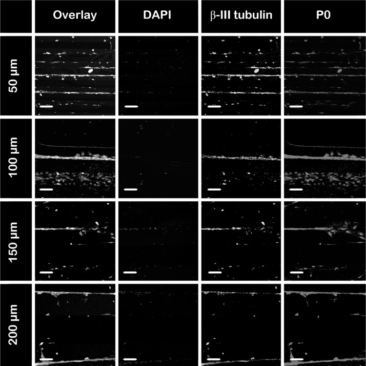
Fig. 6.
Co-localization of SCs with regenerating axons. After 21 days of culture, the co-cultures of DRG neurons with SCs in the different size channels are stained with anti-mouse β-III tubulin (green) to indicate axons, P0 (red) to indicate SCs, and DAPI (blue) to indicate nuclei. First column represents an overlay of all three markers. Fluorescent images are taken with ×20 objective. Scale bar indicates 100 μm. (Color figure online)

