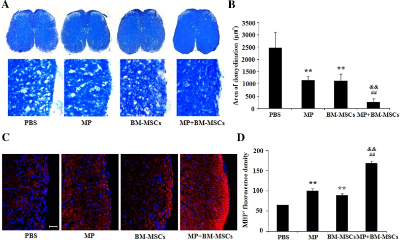
Fig. 3.
Combination treatment with MP and BM-MSCs increases remyelination in EAE spinal cords. EAE mice were sacrificed on day 30 after immunization. Staining of the lumbar spinal cords from each group was performed to detect demyelination and remyelination. A Representative image of spinal cord sections stained with LFB. Magnification: ×400. B Demyelinated areas were quantified using MetaMorph image analysis. Columns, mean; bars, SD. C Representative image of sections immunostained with MBP antibody. Scale bar, 25 μm. D The fluorescence density of staining with MBP antibody was quantified using MetaMorph image analysis. Columns, mean; bars, SD. ** p < 0.01 compared to PBS treatment group; ## p < 0.01 compared to MP treatment group; && p < 0.01 compared to BM-MSCs treatment group; One-way ANOVA with LSD post hoc test. The results are representative of 3 independent experiments

