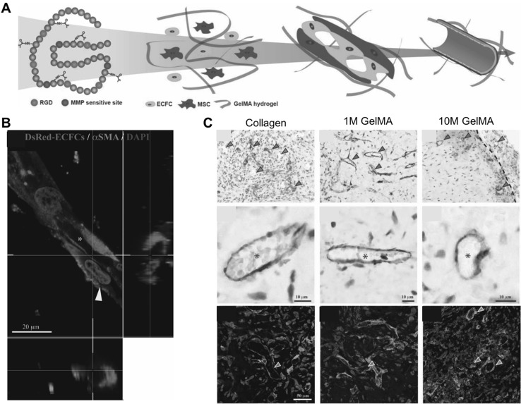
Fig. 2.
Engineered vascular constructs using photo-curable GelMA hydrogels. A Schematic representation of the stepwise process of endothelial lumen formation in the engineered microenvironment. B Premature vessel formation of ECFCs surrounded by α-smooth muscle actin (α-SMA)-expressing MSCs (yellow arrow). Scale bar is 20 µm. C Functional vascular formation in vivo. Immunohistochemistry exhibited that the engineered vasculatures were positively stained for human CD31 (red arrow) and murine capillaries (green arrow), carrying murine erythrocytes (asterisks). Fluorescence images show sections stained with rhodamine-conjugated UEA-1 lectin (to mark human ECFC-lined vessels) and fluorescein isothiocyanate-conjugated anti-α-SMA (to mark perivascular cells; red arrowheads). UEA-1 lectin did not bind to the murine vessels (green arrow). Scale bars are 10 µm and 50 µm
(adapted with permission from Ref [53]). (Color figure online)

