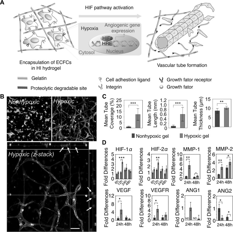
Fig. 3.
Hypoxia-inducible hydrogels to create engineered vasculatures. A Schematic diagram of vascular morphogenesis of ECFCs within artificial hypoxic microenvironment. B Confocal microscopic images of ECFCs cultured within non-hypoxic gels (NG) and hypoxic gels (HG); confocal Z-stacks and orthogonal sections exhibited lumen formation (yellow arrow). Scale bar is 50 µm. C Quantification of vascular tube formation (mean tube coverage, tube length, and tube thickness). D Real-time reverse-transcription polymerase chain reaction for gene expression of ECFCs cultured within two types of hydrogels (NG vs. HG), which is relevant to vascular morphogenesis. Results in C and D are shown as the average value ± s.d. Significance levels were set as *p < 0.05, **p < 0.01, and ***p < 0.001
(adapted with permission from Ref. [16]). (Color figure online)

