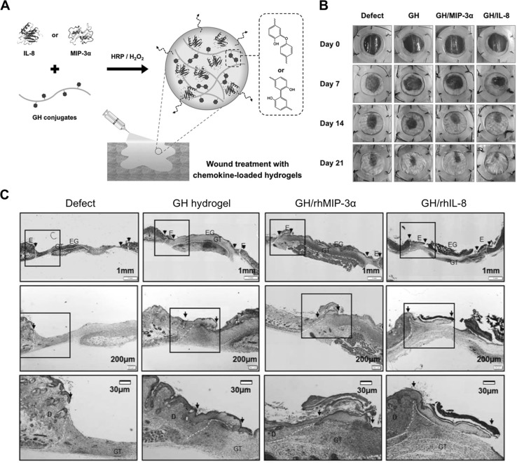
Fig. 5.
Chemokine-loaded in situ forming GH hydrogels for enhanced wound healing in diabetic mouse models. A Schematic illustration of in situ GH hydrogel formation through HRP-mediated cross-linking reaction, encapsulating cell-recruiting chemokines (IL-8 or MIP-3α). B Representative digital images depicting wound healing by the chemokine-loaded GH hydrogels in streptozotocin-induced diabetic mice on day 0, 7, 14, and 21. C Re-epithelialization of the regenerative tissues on day 7 after hydrogel treatment. The explants were subjected to Masson’s trichrome staining and showed re-epithelialization of the damaged tissues with the hydrogels. The arrows indicate regenerated edges of the skin wound. D dermis; E epidermis; EG epithelial gap; GT granulation tissue
(adapted with permission from Ref. [73])

