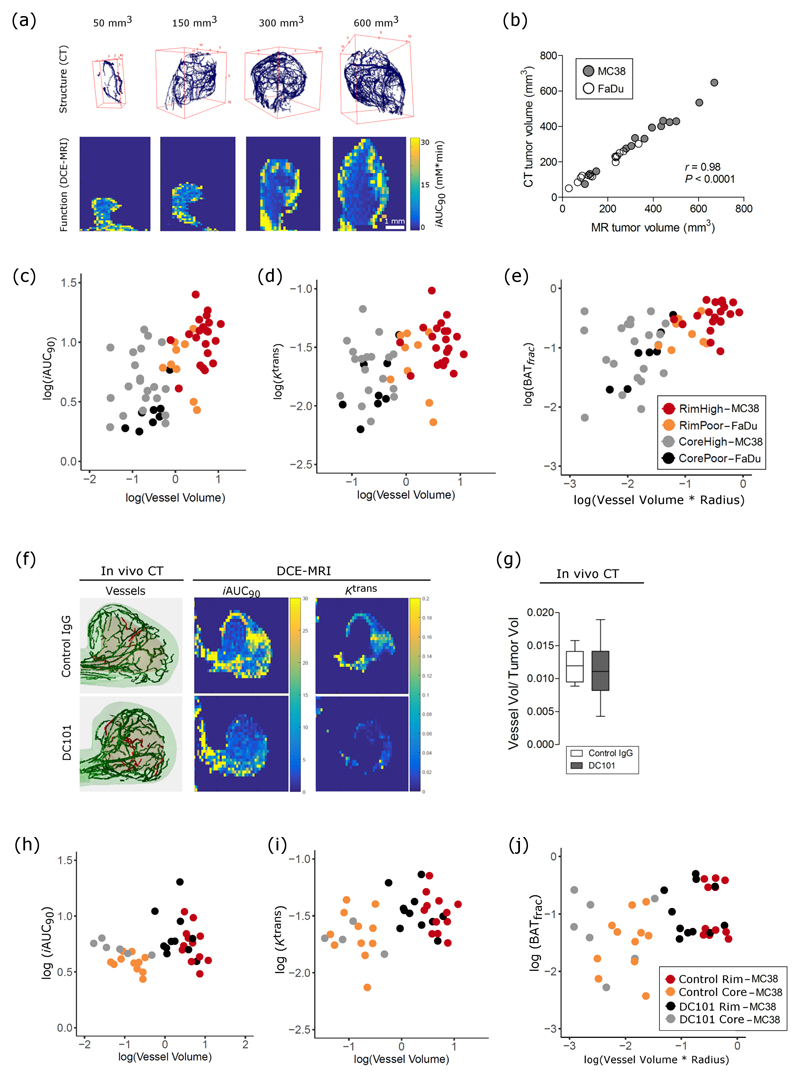
Figure 3.
Functional parameters of tumor perfusion reflect changes in vessel morphology in untreated preclinical tumors in vivo and have an altered relationship to vessel morphology after antiangiogenic treatment. (a) Representative images of segmented vessels from CT and corresponding images of perfusion (iAUC) from a range of tumor volumes. (b) Pearson correlation between analyzed tumor volume on CT and MRI images. (c-e) Relationship of MR parameters, iAUC, Ktrans, and BATfrac to morphological parameters (vessel volume, tortuosity, radius), tumor region (core vs rim), and tumor type (MC38 ‘high’ vs FaDu ‘poor’ vascularization), as identified by linear model analysis. (f) Representative images of segmented vessels from CT and of MR images of iAUC (mM * min) and Ktrans (mL * g-1 * min-1) from control and DC101-treated MC38 tumors. Green vessels are in the rim, while red vessels are in the core. (g) Normalized vessel volume, as measured by CT, does not change with DC101 treatment. Box-whisker plots show median and percentiles (25th and 75th percentile) as boxes, and minimum and maximum values as whiskers. (h-j) Relationship of iAUC and BATfrac, but not Ktrans, with morphological parameters is altered by DC101 treatment in MC38 tumors, as identified by linear model analysis.

