
Figure 2. NOTCH activity, proliferation, cisplatin viability, apoptosis assay and cell cycle analysis of si-NOTCH4 cells.
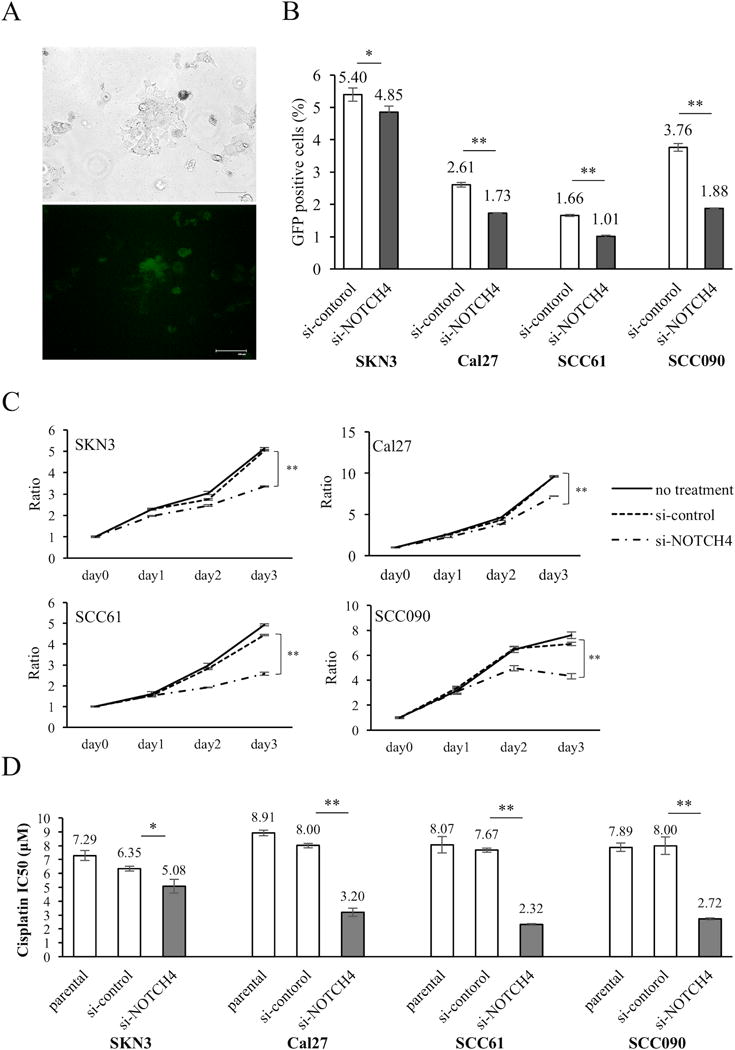
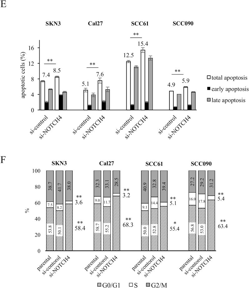
(A) pGreenFire1-Notch plasmid vector was transfected to SKN3 cell. Scale bar indicates 100 μm. (B) NOTCH activity assay of si-control and si-NOTCH4 cells. GFP positive cells have high NOTCH activity. (C) Proliferation assays. si-NOTCH4 cells are compared cell growths to si-control cells on day 3. (D) IC50 of cisplatin in parental, si-control and si-NOTCH4 cells. The IC50 differences between si-control and si-NOTCH4 cells are compared. (E) Apoptosis assays. Total apoptotic fraction is defined as the sum of early and late apoptosis cells. (F) Cell cycle analysis. Each cell cycle phase is compared between si-control and si-NOTCH4 cells. P value is calculated by using Student’s t-test. *: P < 0.05, **: P < 0.01.