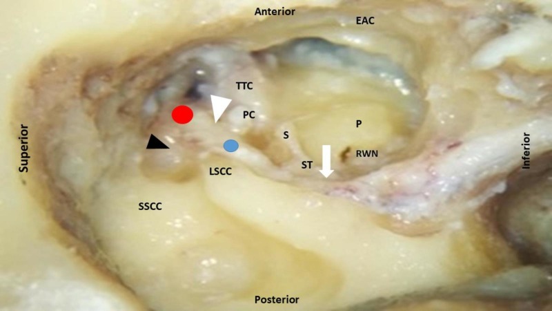
Figure 2. Right temporal bone showing labyrinthine (white arrow head), tympanic (black arrow head) and mastoid segment (white arrow) of the facial nerve. Red dot indicates first genu or geniculate ganglion and blue dot indicates second genu.
EAC: External auditory canal (anterior wall); LSCC: Lateral semicircular canal; P: Promontory; PC: Processus cochleariformis; PSCC: Posterior semicircular canal; RWN: Round window niche; S: Stapes; ST: Stapedius tendon; TTC: Tensor tympani canal; SSCC: Superior semicircular canal.

