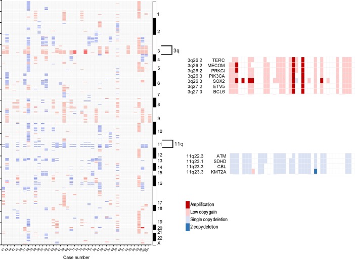
Figure 5. Copy-number alterations in metastatic HPV-associated oropharyngeal cancer.
Copy-number alterations among metastatic HPV+ oropharyngeal tumors (n = 42) arranged by chromosomal band loci (left). Each column represents an individual tumor and corresponding chromosomal gene loci are arranged from top to bottom. Color shading indicates areas of amplification (red) or low copy gain (pink) versus single (light blue) and 2-copy (dark blue) gene deletion. Shown to the right in more detail are regions of recurrent alterations in 3q and 11q with their corresponding gene and genetic loci depicted.

