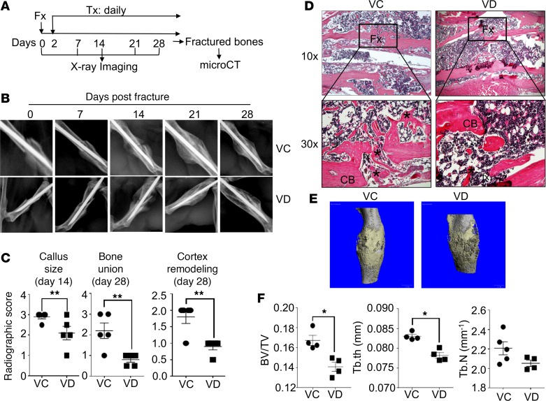
Figure 1. Local s.c. treatment with 1,25(OH)2D during proinflammatory stage impaired fracture healing.
(A and B) B6 mice were subjected to fracture surgery (Fx). Two days later, the animals received a daily s.c. dose of either vehicle control (VC) or 100 ng/kg 1,25(OH)2D (VD) at fracture sites (Tx). X-ray images of the fractured bones were taken at days 0, 7, 14, 21, and 28. Additionally, at day 28, the fractured bones were collected from the animals for μCT analysis. Representative X-ray images are shown. (C) X-ray images from day 14 were quantified for callus size and those from day 28 for bone union and cortex remodeling. **P < 0.01, t test, n = 5. (D) Representative images of H&E staining of the fractured bones are shown. Upper panels, 10×; lower panel:, 30×. Fx, fracture sites; CB, cortical bones; *, new bones. (E) Representative μCT 3-D images are shown. (F) Cumulative data show bone volume/total volume (BV/TV), trabecular thickness (Tb.th), and trabecular number (Tb.N) from the μCT analysis. *P < 0.05, t test, n = 5.

