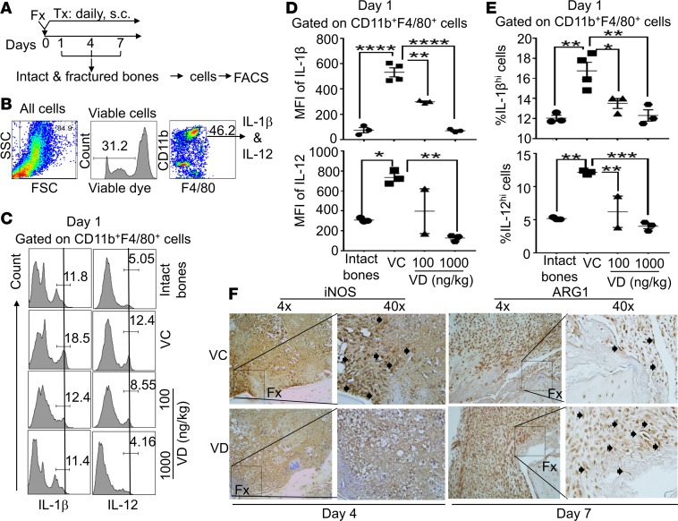
Figure 3. Local s.c. treatment with 1,25(OH)2D at the proinflammatory stage suppressed M1 macrophage differentiation but augmented M2 macrophage differentiation at fracture sites.
(A) B6 mice received femoral fracture surgery (Fx). Immediately after the fracture surgery, the animals received one of the following daily s.c. treatments (Tx) at fracture sites: a) vehicle control (VC), b) 100 ng/kg/mouse 1,25 (OH)2D (VD ), or c) 1,000 ng/kg/mouse 1,25(OH)2D (VD). Additionally, a group of normal healthy mice was included as a control (intact bones). At days 1, 4, and 7 after the treatments, intact and fractured bones were collected, and cells were isolated as described in Methods. The cells were then analyzed by FACS. (B) Gating strategy shows that the cells are gated on viable CD11b+/F4/80+ macrophages for the FACS analysis. (C) Representative FACS plots show the expression of IL-1β (left panel) and IL-12 (right panel) in CD11b+F4/80+ macrophages at day 1 after the treatments. (D) Cumulative data of the mean fluorescent intensities (MFIs) of IL-1β (upper panels) and IL-12 (lower panels) expressions in CD11b+F4/80+ macrophages at day 1. (E) Cumulative data of the percent of IL-1βhi and IL-12hi cells among CD11b+F4/80+ macrophages at day 1. *P < 0.05, **P < 0.01, ***P < 0.001, ****P < 0.0001, ANOVA test, n = 3. (F) B6 mice were subjected to fracture surgery and, immediately after the fracture surgery, received a dose of either VC or 100 ng/kg 1,25(OH)2D (VD). At days 4 and 7, fractured bones were collected. Paraffin embedded sections from day 4 were stained for iNOS and those from day 7 were stained for ARG1 by 3,3′-Diaminobenzidine (DAB) staining. Arrows indicate positively stained cells. Representative images are shown.

