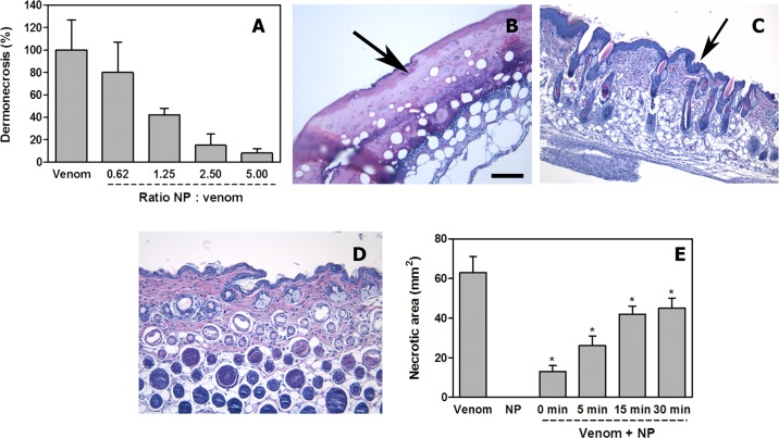
Fig 6. Inhibition of dermonecrotic activity of N. nigricollis venom by NPs.
(A) A fixed amount of venom was incubated with variable amounts of NPs to attain several ratios. Controls included venom incubated with saline solution instead of NPs. Upon incubation, aliquots of the mixtures, containing 100 μg venom, were injected intradermally in mice. Seventy-two hours after injection, the areas of necrotic lesions in the inner side of the skin were measured. Necrosis was expressed as percentage, 100% corresponding to the necrotic area (60 mm2) in mice receiving venom alone. NPs inhibited, dose-dependently, the dermonecrotizing activity of the venom. (B, C, D) Light micrographs of sections of the skin of mice 72 h after injection of 100 μg N. nigricollis venom incubated with saline solution (B), with NPs at a NP: venom (w : w) ratio of 5.0 (C), or after injection of NPs alone (D). In (B) there is ulceration, with loss of epidermis and the formation of a hyaline proteinaceous material (arrow); skin appendages are absent and there is a prominent inflammatory infiltrate. In (C) NPs inhibited the ulcerative effect, evidenced by the presence of epidermis (arrow) and skin appendages, whereas an inflammatory infiltrate is observed in the dermis. Skin injected with NPs alone (D) shows a normal histological pattern. Hematoxylin-eosin staining. Bar represents 100 μm. (E) Inhibition of dermonecrosis by N. nigricollis venom in experiments involving intradermal injection of 100 μg venom followed by the injection of NP suspension (5.5 mg/mL), in the same region of venom injection, at various time intervals. A significant reduction in the extent of dermonecrosis was observed (* p < 0.05), although the extent of inhibition decreased with time. NP alone did not induce skin damage.

