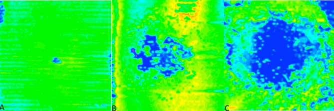
Fig 2. Comparison of EZ loss.
(A) En-face map of the EZ layer with minimal loss, as shown by the majority of the green, from a patient included in this normative EZ study. (B) En-face map with moderate EZ loss, with more central blue, from a patient with Stargardt disease. (C) Severe loss, as shown by the expansive central blue areas, also from a patient with Stargardt disease.

