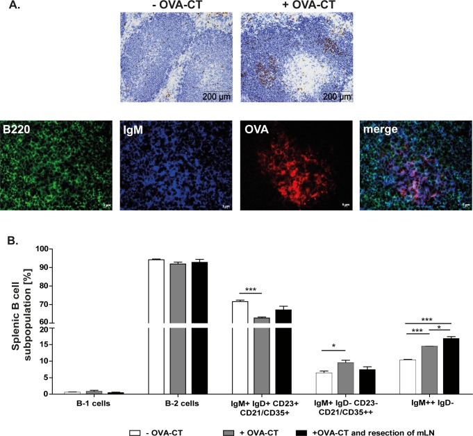
Fig 2. Oral application of OVA-CT induces the proliferation of splenic memory IgM+ and marginal zone B cells.
Control and mLN resected mice (n = 3–4) were orally challenged with OVA-CT. 25 days after the challenge the spleen was removed and analysed. A. Representative immunohistological staining of cryosections showing incorporated BrdU (brown) and B cells (blue) in the spleen of control mice +/- OVA-CT administration. Focusing on germinal centers after OVA-CT challenge, antigen specific B cells were stained using Alexa Fluor 555 conjugated OVA (red), IgM (blue) and B220 (green). B. B1 and B2 cells were distinguished by their IgM, CD19, CD5 and CD11b expression. In addition, B2 cells were separated into follicular B cells (IgM+ IgD+ CD21/35+ CD23+), marginal zone B cells (IgM+ IgD- CD21/35++ CD23-) and memory B cells and marginal zone B cells (IgM++ IgD-). Significant differences in the unpaired t-test are indicated by *P< 0.05; ***P< 0.001.

