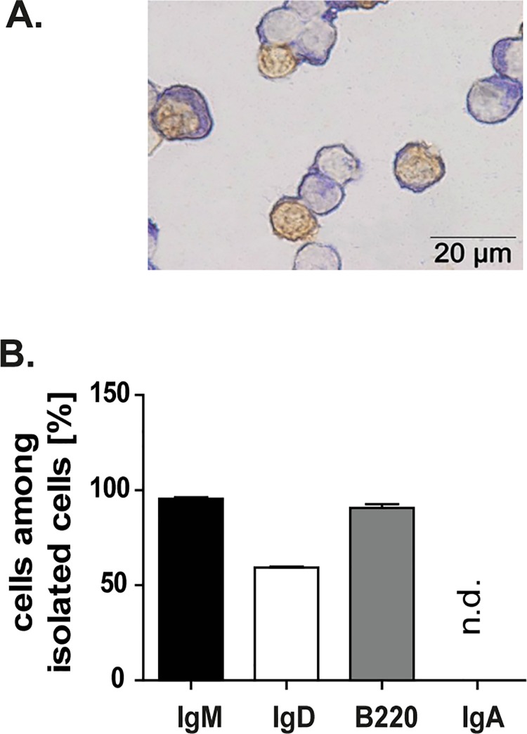
Fig 3. Splenic IgM+ B cells proliferate after orally applied antigen challenge.
A. Splenic IgM+ B cells of C57BL/6-Ly5.1 mice were isolated using the MACS technique and cytospins of isolated cells were performed. The illustration shows the IgM+ B cells in blue and the proliferating IgM+ B cells (BrdU+ cells) in brown. The purity of the isolated cells was near 100%. B. Flow cytometry analysis of isolated cells. Majority of isolated cells were IgM+ B cells. IgA+ B cells were not detected (n.d). Means and standard error of the mean are given for seven independent experiments.

