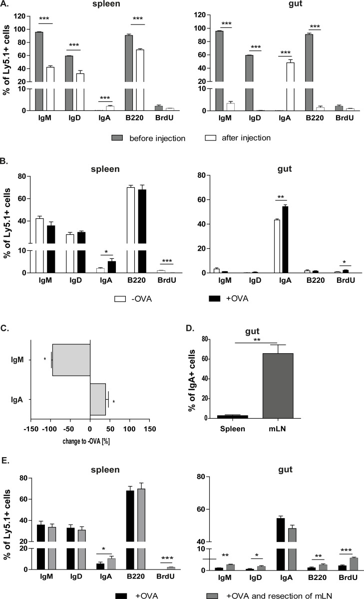
Fig 5. IgM+ B cells that migrate into the gut are OVA-specific IgA+ plasma cells.
The mLN resection increases IgA expression in the spleen, but not in the gut. Isolated OVA-CT treated IgM+ C57BL/6-Ly.5.1 cells were analysed for their immunoglobulin expression by flow cytometry. A. Comparison of isolated cells before and after the transfer into recipients. After migration of the injected cells into the spleen and the gut percentages were compared to the initial cell population. B. IgM+ B cells were injected into non-treated (-OVA) and treated (+OVA) mice and the percentage of different markers expressed on these cells after migrating in the spleen and the gut were analysed using flow cytometry. Significant differences in the unpaired t-test are indicated by *P< 0.05; **P< 0.01; ***P< 0.001. C. The serum of non-treated (-OVA) and treated (+OVA) animals, which received splenic IgM+ B cells, was tested in ELISA for OVA-specific antibodies. OVA-specific antibodies of non-treated animals are equalized as 100%. The measured OVA-specific antibodies of treated animals are calculated as a % change to non-treated (-OVA) mice (x-axis). Means and standard error are given from 3–4 independent experiments (significant differences in the unpaired t-test are indicated by *: P < 0.05). D. IgM+ B cells of OVA/CT treated mice were isolated from the spleen or the mLN and co-transferred to WT mice. After OVA treatment intestines of these mice were analysed and IgA+ B cells from the mLN and spleen were measured. Two independent experiments were performed (n = 4). Significant differences in the paired t-test are indicated by **P< 0.01. E. Recipients underwent the mLN resection. After splenic IgM+ B cell transfer recipients were orally challenged with OVA. Four independent experiments were performed. Significant differences in the unpaired t-test are indicated by *P< 0.05; **P< 0.01; ***P< 0.001.

