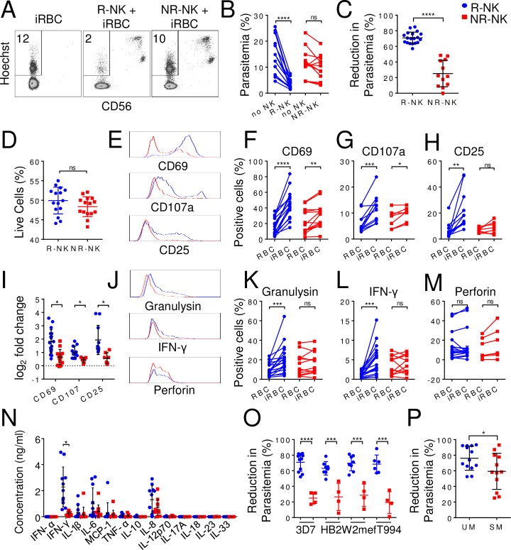
Fig 1. Variation of NK cell responses to iRBCs among different individuals.
Human NK cells were purified (>95%) from fresh blood and co-cultured with iRBCs for 96 h. Parasitemia and the expression of various NK cell surface markers and intracellular proteins were assayed by flow cytometry. The level of cytokines in the culture supernatants was quantified by multiplex immunoassay at 48 h. (A) Representative CD56 vs. Hoechst staining profiles of iRBCs alone, iRBCs co-cultured with responder NK cells (R-NK) or with non-responder NK cells (NR-NK). The number represents the percentage of iRBCs (parasitemia). CD56-positive cells are NK cells. (B) Paired plots of parasitemia in the absence of NK cells (No NK) and with either R-NK or NR-NK. (C) Comparison of reduction in parasitemia in the presence of either R-NK (blue) or NR-NK (red) cells from different malaria-naïve individuals. (D) Percentage of live (DAPI-negative) NK cells after 96 hrs of co-culture. (E) Representative histograms comparing CD69, CD107 and CD25 expression on R-NK and NR-NK cells from one responder (blue trace) and one non-responder (red trace). (F-H) Paired plots showing changes in the percentage of NK cells positive for CD69 (F), CD107a (G) and CD25 (H) following co-culture with either RBC or iRBC. (I) Comparison of log2 fold change in expression levels of CD69, CD107 and CD25 between R-NK and NR-NK cells following co-culture with iRBC. (J) Representative histograms comparing granulysin, IFN-γ and perforin expression on R-NK and NR-NK cells from one responder and one non-responder. (K-M) Paired plots showing changes in the percentage of NK cells positive for granulysin (K), IFN-γ (L) and perforin (M) following co-culture with either RBC or iRBC. (N) Comparison of soluble mediators in the culture supernatants. (O) Comparison of reduction in parasitemia following co-culture of R-NK or NR-NK cells in the presence of different strains of parasites. R-NK and NR-NK cells were co-cultured with the indicated parasite strain for 96 h. Parasitemia was quantified by flow cytometry. (P) Comparison of reduction in parasitemia in the presence of NK cells from either uncomplicated malaria (UM) patients or severe malaria SM patients. NK cells were purified from frozen buffy coat and co-cultured with 3D7-infected RBCs for 96 h and parasitemia was quantified by flow cytometry. Each symbol in B-D, F-I, K-P represents a different individual. Joined lines show experimental pair. Error bars represent mean ± SD. * p<0.05, ** p<0.01, *** p<0.001, **** p<0.0001, ns: not significant.

