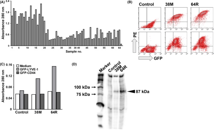
Figure 1.

Production and characterization of anti‐mouse lymphatic vessel endothelial hyaluronan receptor 1 (LYVE‐1) rat monoclonal antibodies (mAb). A, First screening: 64 hybridoma clones with strong reactivity to soluble LYVE‐1 proteins fused to GFP on ELISA. Samples 38 (38M) and 64 (64R) are the clones selected in the 2nd screening. Sample 65, control (right end, mAb‐). B, Second screening: flow cytometry analysis of the 38M and 64R mAb against GFP‐LYVE‐1‐expressing HEK293F (upper) and RH7777 (lower). PE, phycoerythrin. C, Reactivity of the 38M and 64R mAb with serum‐free culture supernatants from HEK293F cells transfected with GFP‐LYVE‐1 or GFP‐CD44 on ELISA. D, SDS‐PAGE of HEK293F cells transfected with GFP‐LYVE‐1, and surface‐biotinylated and immunoprecipitated with anti‐mouse LYVE‐1 rat mAb. Proteins were visualized with Elite ABC and peroxidase substrates. Arrowheads, positions of mAb‐bound GFP‐mouse LYVE‐1 proteins
