
Figure 5.
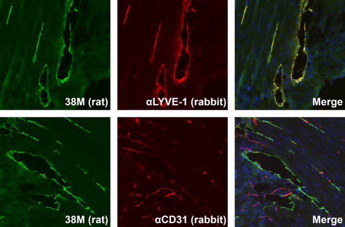
Confocal analysis of lymphatic vessel endothelial hyaluronan receptor 1 (LYVE‐1) in mouse stomach tissue. Paraformaldehyde (PFA)‐fixed mouse tissue sections were treated with the 38M rat mAb in combination with anti‐LYVE‐1 or anti‐CD31 rabbit antibodies overnight at room temperature. Tissue sections were treated with species‐specific Alexa Fluor 488‐conjugated anti‐rat and Alexa Fluor 678‐conjugated anti‐rabbit IgG for 60 minutes at room temperature. The localization of antibody‐defined components (LYVE‐1 and CD31) was observed using a confocal microscope