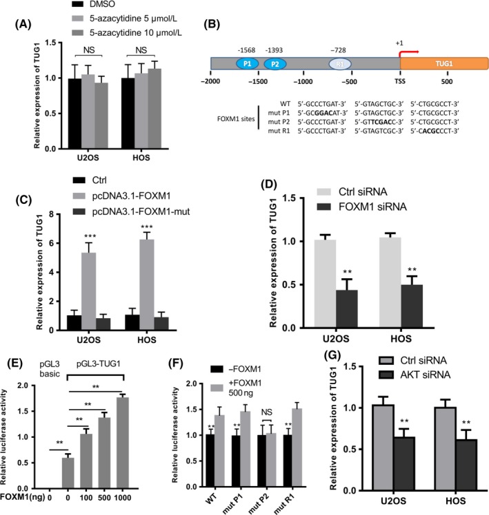
Figure 2.

Identification of TUG1 (Taurine Upregulated Gene 1) in an protein kinase B / Forkhead Box M1 (AKT/FOXM1) axis regulated in osteosarcoma cells. (A) Quantitative real‐time PCR analysis of TUG1 in U2OS and HOS treated with DMSO or 5‐azacytidine (5 μmol/L or 10 μmol/L) for 48 h (n = 3). (B) A schematic illustration of the TUG1 promoter region. The wild‐type and mutant sequences of two predicted binding sites, P1 (‐1568) and P2 (‐1393), and one random site, R1 (‐728), are underlined. (C) Quantitative real‐time PCR analysis of TUG1 in U2OS and HOS cells transfected with 500 ng indicated plasmids after 48 h (n = 3). (D) Quantitative real‐time PCR analysis of TUG1 in U2OS and HOS cells after transfection with control or FOXM1 siRNA (n = 3). (E) A combination of 500 ng pGL3‐TUG1 (or pGL3‐Basic as a negative control), 50 ng pRL‐TK and an increasing number of pcDNA3.1‐FOXM1 plasmids were co‐transfected into U2OS cells. Luciferase activity was tested after 48 h (n = 3). pGL3‐basic was used as a negative control. (F) A combination of 500 ng pGL3‐TUG1 promoter carrying either wild‐type sequence or mutations in two putative FOXM1 binding sites and one random site, 50 ng pRL‐TK and 500 ng pcDNA3.1‐FOXM1 were co‐transfected in U2OS cells. Luciferase activity was tested after 48 h (n = 3). (G) The expression levels of TUG1 in U2OS and HOS cells transfected with control or si‐AKT for 48 h (n = 3). Error bars indicate the mean ± SD. **P < .001, ***P < .0001
