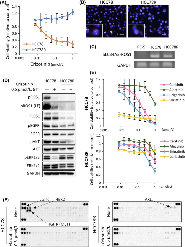
Figure 1.

Establishment of the crizotinib‐resistant cell line, HCC78R. A, Cell proliferation assay of HCC78 and HCC78R cells treated with the indicated concentrations of crizotinib. Error bars: SD. All experiments were performed in triplicate. B, FISH analysis of ROS1 fusion gene in HCC78 and HCC78R cells. Red probes hybridized to the 5′ region of ROS1 and green probes to the 3′ region. In the presence of ROS1 rearrangement, the 2 colors are observed separately. HCC78R cells maintained ROS1 fusion genes (98% ROS1 fusion FISH positive) to the same extent as the parental HCC78 cells (94% ROS1 fusion FISH positive) C, Detection of SLC34A2‐ROS1. Complementary DNA (cDNA) derived from PC‐9 (negative control), HCC78 and HCC78R were examined by PCR. D,F. Immunoblots and phospho‐receptor tyrosine kinase (RTK) arrays in HCC78 and HCC78R cells. These cells were cultured in normal medium for 4 d, after which both types of cells were exposed to .5 μmol/L crizotinib for 6 h. E, Cell proliferation assay of HCC78 and HCC78R cells treated with the indicated concentrations of ALK/ROS1 inhibitors, ceritinib, alectinib, brigatinib and lorlatinib. Error bars: SD. All experiments were performed in triplicate
