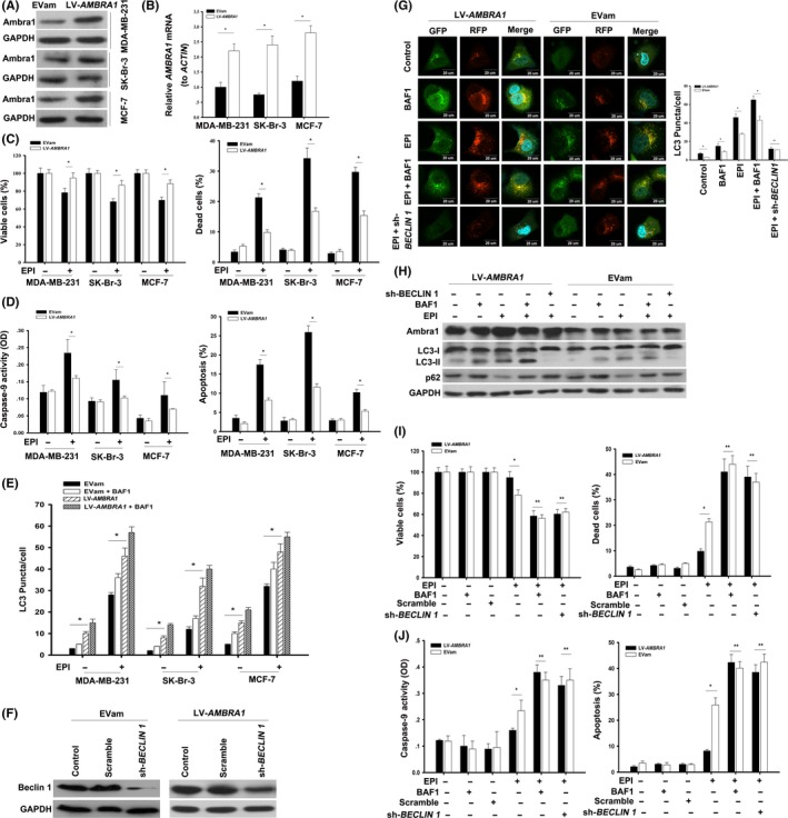
Figure 2.

Overexpression of Ambra1 increased the resistance of breast cancer cells to epirubicin (EPI). A, MCF‐7, MDA‐MB‐231 and SK‐Br‐3 cells were transfected with LV‐AMBRA1 or empty vector (EVam) for 48 h, and the protein of Ambra1 was tested by western blotting. Meanwhile, the mRNA of AMBRA1 was analyzed by quantitative RT‐PCR (B). The results (mean ± SE) are from 3 independent experiments (*P < 0.05). C, Cells incubated with LV‐AMBRA1 or EVam for 48 h, following treatment with 2.2 μmol/L EPI for 24 h; then, cell viability and mortality were analyzed. Moreover, caspase‐9 activity and apoptosis were assayed (D). The results (mean ± SE) are from 3 independent experiments (*P < 0.05). E, Cells expressing RFP‐GFP‐LC3 were incubated with LV‐AMBRA1 or EVam for 48 h. After that, these cells were treated with 2.2 μmol/L EPI for 24 h in the presence or absence of Bafilomycin A1 (BAF1, 20 nmol/L). Autophagy was assessed with the LC3 puncta. The results (mean ± SE) are from 3 independent experiments (*P < 0.05). F, MDA‐MB‐231 cells were transfected with LV‐AMBRA1 or EVam for 48 h and then incubated with scrambled shRNA or sh‐BECLIN 1 for additional 48 h. Then, the protein of Beclin 1 was detected by western blotting. G, MDA‐MB‐231 cells expressing RFP‐GFP‐LC3 were incubated with LV‐AMBRA1 or EVam for 48 h. Then, the cells were transfected with scrambled shRNA or sh‐BECLIN 1 for 48 h, followed by treatment with 2.2 μmol/L EPI for another 24 h in the presence or absence of Bafilomycin A1 (BAF1, 20 nmol/L). After treatment, the fluorescence of RFP and GFP was observed with a fluorescence microscope (600×) and LC3 puncta were counted. The nucleus was stained with Hoechst. The results (mean ± SE) are from 3 independent experiments (*P < 0.05, **P > 0.05). In parallel, the proteins of Ambra1, LC3‐I/II and p62 were detected by western blotting (H). Then, (I) and (J), cell viability, cell death, caspase‐9 activity and apoptosis were analyzed. The results (mean ± SE) are from 3 independent experiments (*P < 0.05, **P > 0.05)
