
Figure 6.
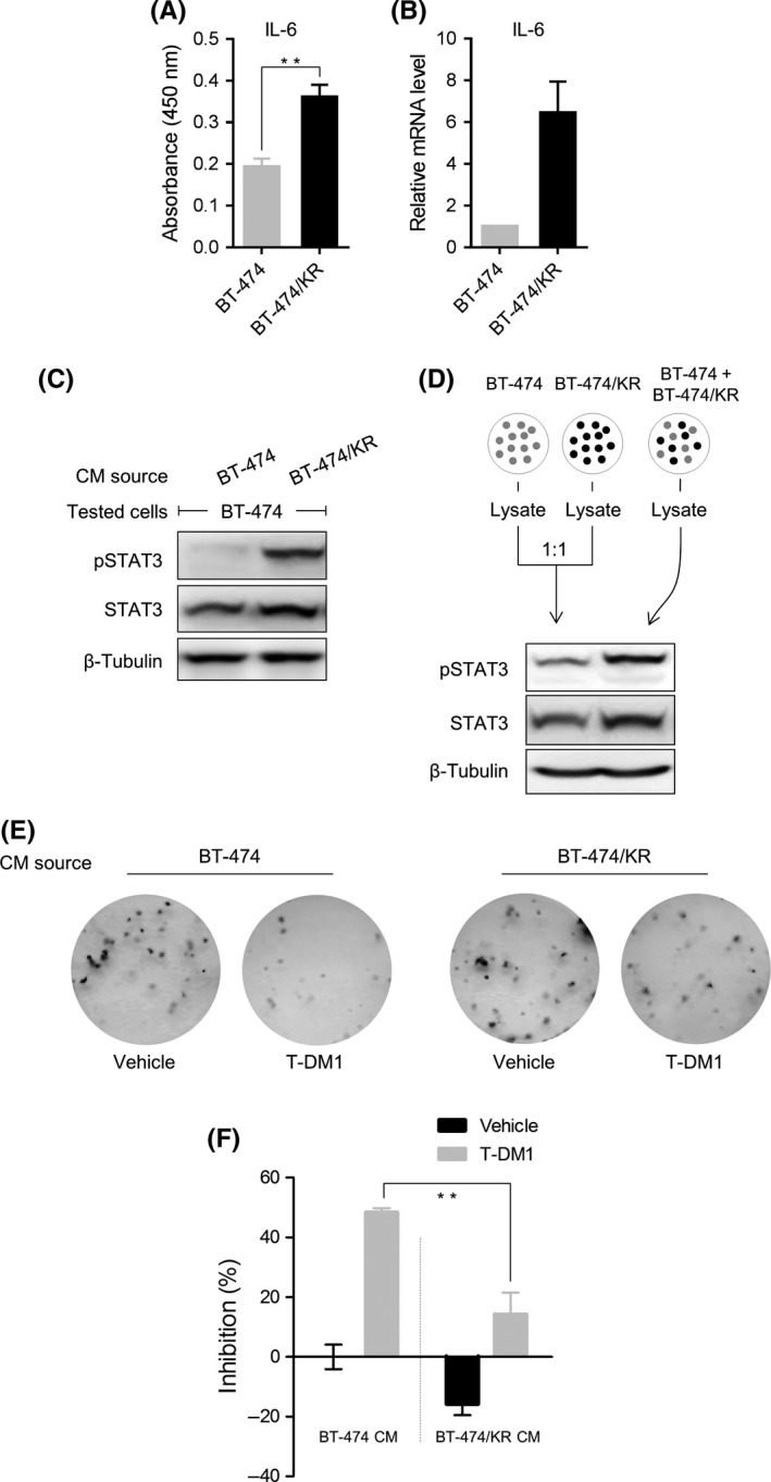
Factors secreted by BT‐474/KR cells confer resistance on BT‐474 cells. A, BT‐474 and BT‐474/KR cells were cultured for 96 h. Interleukin‐6 (IL‐6) level in the supernatants was analyzed on ELISA. B, mRNA level of IL‐6 in BT‐474 and BT‐474/KR cells was analyzed on quantitative reverse transcription‐polymerase chain reaction. C, BT‐474 cells were incubated with conditioned media (CM) from BT‐474 or BT‐474/KR cells for 24 h. Signal transducer and activator of transcription 3 (STAT3) activation was measured on western blotting. D, BT‐474 and BT‐474/KR cells were cultured separately, and lysates were mixed at a 1:1 ratio, or BT‐474 and BT‐474/KR cells were co‐cultured at a 1:1 ratio. STAT3 activation was measured on western blotting. E, BT‐474 cells were incubated in CM from BT‐474 or BT‐474/KR cells, with or without trastuzumab‐emtansine (T‐DM1; 30 ng/mL). Colonies were stained with crystal violet. F, The number of colonies formed in (E) was quantified; percent inhibition is expressed relative to colonies formed following incubation in CM from BT‐474 cells without T‐DM1 exposure. Error bars represent mean ± SD from triplicates. **P < .01