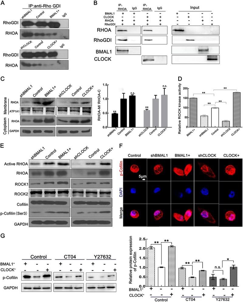
Fig. 5. CLOCK and BMAL1 regulate the RHOA-ROCK-CFL pathway.
a The complex was immunoprecipitated from total protein extracts collected from HeLa cells transfected with corresponding plasmids. The proteins were then analyzed by Co-IP with antibodies against RhoGDI. b The TNT® Quick Coupled Transcription/Translation System was used here for protein (BMAL1, CLOCK, RHOA, and RHOGDI) synthesis. The synthesized proteins were then analyzed by Co-IP with corresponding antibodies. c The RHOA distribution was affected by BMAL1 or CLOCK, as detected by a cytosolic/membrane fractionation assay in HeLa cells. *p < 0.05; **p < 0.01, n.s. no significant. d HeLa cells transfected with the indicated vectors were harvested and analyzed using a 96-well ROCK Activity Assay Kit. The activity levels were normalized to those in the control, which was given an arbitrary value of 1 **p < 0.01. e CLOCK and BMAL1 can promote the activation of RHOA and the expression of RHOA and other proteins involved in the RHOA-ROCK-CFL pathway. Activated RHOA was pulled down using a GST-Rhotekin-Rho-binding domain (RBD) fusion protein. f Immunofluorescent staining of p-cofilin (Ser-3) in HeLa cells transfected with BMAL1+, CLOCK+, shBMAL1, and shCLOCK. Scale bar, 5 µm. g HeLa cells transfected with the indicated plasmids were treated with 1 μg/ml CT04 (RHOA inhibitor) for 2 h and Y27632 (ROCK inhibitor) for 20 h before whole-cell lysates were extracted and analyzed. Statistical results of the p-cofilin protein level are shown to the right *p < 0.05; **p < 0.01, n.s. no significant

