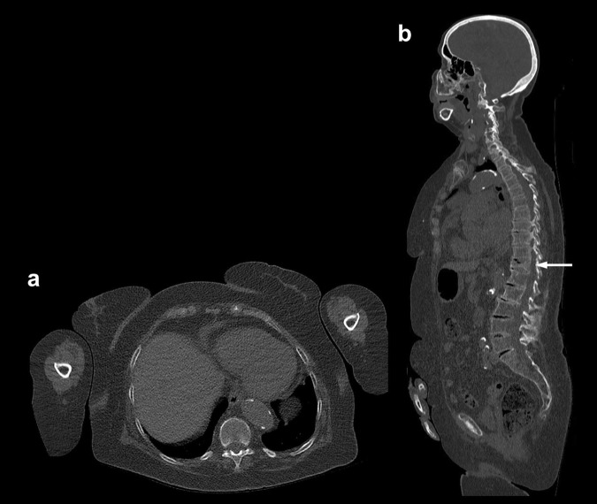
Fig. 2.
WBLDCT scan of the same patient as in Fig. 1, performed on a 128-slice CT scanner three years later with the same tube voltage and time-current product (120 kV, 60 mAs). The arms are lying more anteriorly than in Fig. 1, and an iterative reconstruction algorithm was used to reduce artifacts and image noise. a Axial slice (2/1 mm thickness/increment) at the level of the T10 vertebral body and b Sagittal MPR image. Overall image quality is significantly improved compared to the images of Fig. 1. The trabecular structure of the T10 vertebral body is readily appreciated in a and spinal anatomy is well depicted on b. Note new compressive vertebral fracture of T11 (arrow in b). MPR multiplanar reformation

