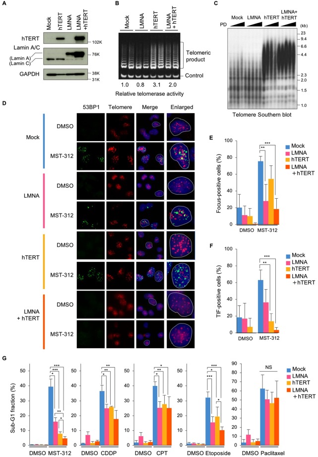
Figure 6.
Telomere elongation and lamin A expression confers resistance to MST-312. (A,B) Telomere elongation and lamin A expression in NCI-H522 cells. Cells were infected with hTERT and/or LMNA expressing retrovirus. Cells were subjected to western blot analysis for detection of hTERT and lamin A expression (A) and TRAP assay for detection of telomerase activity (B). For (A) full-length blots were presented in Supplementary Fig. S5. (C) Telomere southern blot analysis. Genomic DNA was prepared and subjected to southern blot analysis with [32P]-labelled telomeric probe. (D) Telomere immuno-FISH analysis. Cells were treated with 5 μM MST-312 for 48 h and subjected to immuno-FISH analysis. Green: 53BP1; red: telomere DNA; blue: DAPI stain of DNA. (E) Quantitation of DNA damage foci in (D). Cells with more than four 53BP1 foci were counted as positive. (F) Quantitation of telomeric DNA damage (TIF and enlarged TIF) in (D). Cells with more than four TIF were counted as positive. (G) Apoptosis induced by MST-312 and anticancer drugs. Cells were treated with 5 μM MST-312, 3.3 μM cisplatin (CDDP), 10 nM camptothecin (CPT), 1.6 μM etoposide or 5 nM paclitaxel for 96 h. Cells were stained with propidium iodide and the apoptotic sub-G1 fraction of the cell cycle was quantitated by flow cytometry. Error bar indicates standard deviation of four (E,F) or at least three (G) experiments. Statistical significance was evaluated by Tukey-Kramer test. *P < 0.05, **P < 0.01, ***P < 0.001. NS: not significant.

