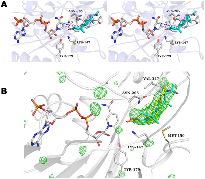
Figure 6.
PmMORV150M progesterone binding. (A) Stereo-view of the active site of PmMORV150M in complex with NADP+ and progesterone with protein and side chains shown in gray. Selected residues near the ligand are shown in sticks. NADP+ is colored by atom with carbons in gray. Progesterone is colored by atom with carbons in cyan. The binding mode of progesterone postulated by Thorn, et al.24 is shown and colored by atom with carbons in gray. Note the steric clash with ASN-205 for this binding mode. Distances between the reactive hydride and carbon-carbon double bond to be reduced are 6.3 Å and 7.1 Å for the PmMOR and Thorn, et al. conformation, respectively. (B) PmMORV150M progesterone omit map contoured at +3.5 standard deviations (σ) above the mean shown in green mesh. Selected side chain residues are drawn as sticks and labeled. The PmMORWT progesterone ligand is shown in yellow sticks and the PmMORV150M progesterone is shown in cyan sticks.

