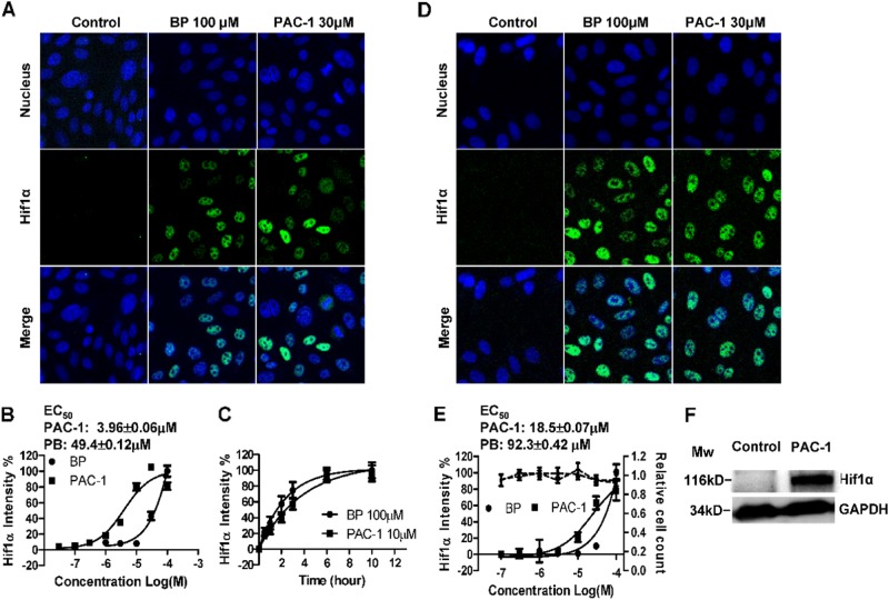
Fig. 2. PAC-1 induces the stabilization of HIF1α under normoxic conditions.
HIF1α-EGFP_CHO cells were treated with the indicated drugs for 3 h before fixation, acquisition of fluorescence images (a), and quantitative analysis (b). c Time course of HIF1α accumulation in HIF1α-EGFP_CHO cells. d–f HepG2 cells were treated with the indicated drugs for 3 h, and HIF1α levels were detected using specific antibodies. d Fluorescence images of stabilized HIF1α in HepG2 cells. e Concentration-dependent accumulation of HIF1α in HepG2 cells (black solid line) and changes in cell counts (black dashed line). f Western blotting of HIF1α accumulation in HepG2 cells. Data represent means ± SEs of three independent experiments

