
Figure 1.
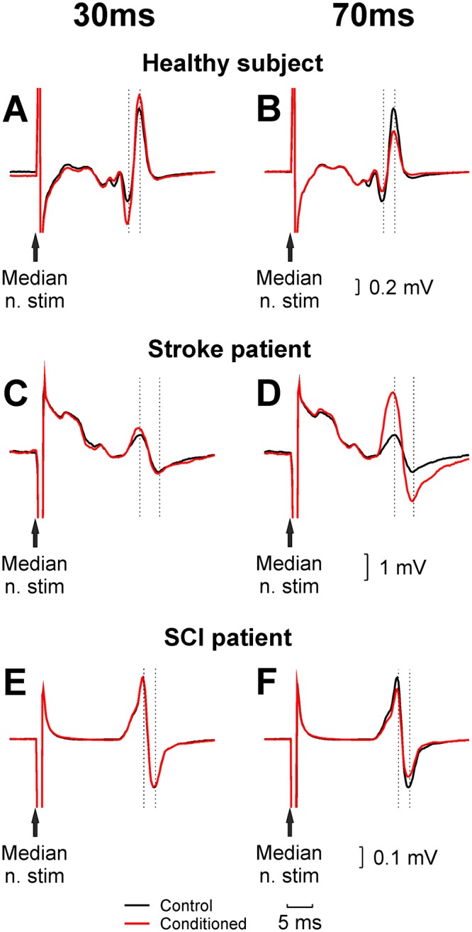
Example H-reflex recordings from the FCR muscle. Each panel shows an unconditioned H-reflex (black), and an H-reflex conditioned by preceding stimulation of the ECR muscle (red). Results are shown for a single healthy subject (A,B) patient with stroke (C,D Ashworth score 2) and spinal cord injury (E,F Ashworth score 0). Left panels (A,C,E) used a conditioning interval of 30 ms, which generates facilitation in healthy subjects; right panels (B,D,F) used an interval of 70 ms, at which the reflex is suppressed in healthy subjects.