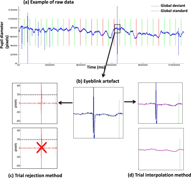
Figure 2.
Raw data and two methods to process eye-blink artefacts (a) Example of raw data (blue curve): temporal evolution of pupil horizontal diameter (in pixels) from the right eye of one participant, superimposed with global standard trials in green and global deviant trials in red. (b) Zoom on a trial with an eye blink artefact processed with two different methods. (c) With the trial rejection method a trial was rejected if the difference between two successive temporal points was larger than 200 pixels. (d) With the trial interpolation method (see details in the text) any identified artefact (blue) was corrected using a cubic interpolation (bottom magenta curve).

