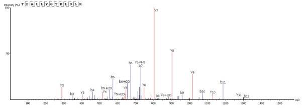
FIGURE 3.

The complete Sarconesin aa sequence was obtained by mass spectrometry (MS/MS) fragmentation; representative de novo sequencing of Sarconesin. CID spectrum from mass/charge (m/z) of its double charged ion gave [M + 2H]2 + , m/z 736.9266. The ions from y (red) and b (blue) series (marked at the top of the spectrum) represent the primary structure: TPm( + 16)LLVGTKLDLR. The sequenced peptide’s internal fragments whose ions were found in the spectrum are represented by standard aa letter code.
