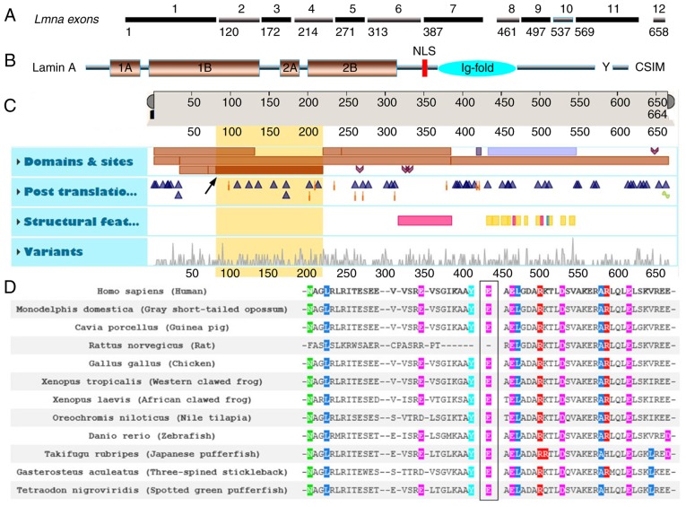
Figure 3.
(A) Sketch map of exons 1–12 of LMNA. (B) Sketch map of lamin A protein. Y represents the site for zinc metallopeptidase STE24 hydrolysis and CSIM represents the target for farnesylation. (C) Features viewer and functional consequence of LMNA p.E82K (GeneView). Yellow region of interest, amino acids 81–218; description of Coil 1B in LMNA and the arrow indicates the p.E82K mutation. (D) Homology comparison of lamin A protein among different species: Amino acid alignment revealed conservation among species for glutamic acid at position 82 in LMNA. CSIM, CaaX motif; LMNA, lamins A/C.

