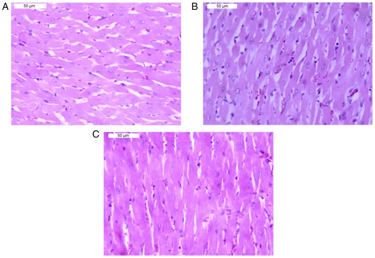
Figure 4.
Morphology of cardiac tissue in rats under an optical microscope. Hematoxylin and eosin staining (magnification, ×200). (A) Control group. (B) Chuan-wu treated group: Veins were dilated and congested, and slight inflammatory cell infiltrations were observed. (C) Ber 16 mg/kg group: A normal myocardium was exhibited.

