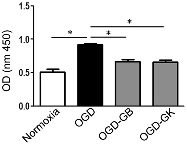
Figure 2.

Effect of GB and GK on PAF inhibition in OGD astrocytes. Primary astrocytes were exposed to OGD for 3 h. At the beginning of re-oxygenation, GB and GK (30 ug/ml, dissolved in DMSO) were added. The same volume of DMSO was added as a control. Following 24 h, the supernatants were collected for the PAF assay. Quantitative results are the mean ± standard error of the mean, and analyzed from three independent experiments with similar results. *P<0.05, as indicated. GB, ginkgolide B; GK, ginkgolide K; OGD, oxygen-glucose deprivation; PAF, platelet-activating factor; OD, optical density; DMSO, dimethyl sulfoxide.
